To speed up medical documentation, many neurologists prefer dictating their notes and outsourcing medical transcription to a reliable vendor. Accuracy is an important element when it comes to transcribing neurology reports. A reliable service provider offers customized solutions to individual neurologists, physicians’ groups, hospitals, neurology clinics, acute care clinics and long-term care clinics. They provide radiology transcription for MRIs, CT scans, X rays etc.
Imaging technologies are used during surgery to guide the movement of surgical instruments. They are also used to deliver treatment to a specific body part without invasive surgery. Neurologists may use MRI scans and CT scans to get a better resolution of neural structures. It helps them to quickly diagnose brain injury, stroke, spinal cord tumours, and infections. Sometimes, imaging studies may be dictated as procedure notes or surgical reports if the study is performed in an operating room. So, it is important to ensure that radiology notes are accurately recorded and transcribed to help neurologists diagnose better and provide the most appropriate treatment as soon as possible.
Let us look at a research study where MRIs had a significant role to play.
Study Reveals Brain Regions that May Control Fatigue
Scientists at John Hopkins Medicine used MRI scans and computer modelling to find out parts of the human brain that can regulate efforts to control fatigue. The results of the study were published in Nature Communication.
For the study, the team developed a new way to quantify how people feel fatigued, which varies from people to people. 20 participants, with an average age of 24 (18 to 34), were asked to make a risk-based decision about exerting a certain amount of physical effort. Nine out of 20 participants were female.
The participants were asked to squeeze a sensor after training them to recognize a scale of effort. For example, zero was equal to no effort and 50 units of effort were equal to half the participant’s maximum force. The participants continued the grip exercise for 17 blocks for 10 trials until they were fatigued. After that they were offered one of two choices for making such an effort – a random choice based on a coin flip (which offered the chance to exert no effort) or a predetermined set effort level. This was with a view to understand how each participant valued their effort. It would also help understand how their brains and minds decided how much effort to exert.
The researchers used computerized programs to know how participants felt about the prospect of exerting particular amounts of effort while they were fatigued. The study found that most of the participants were risk averters. This means that when they were fatigued they were less willing to take the chance of having to exert too much.
Another group of 10 people were trained on gripping system but were not given fatigue trials. The participants’ brain activity was evaluated for the research with functional MRI scans, which track blood flow through the brain and show which neurons are firing most often. Just as in previous findings, it was found that when the participants chose between the two options, brain activity seemed to increase in all participants in the area of the brain known as the insula. MRI scans were also used to examine the motor cortex of the brain (which is responsible for exerting the effort itself) when the participants were fatigued. The research team found that the motor cortex was deactivated when the participants were making a choice. Participants whose motor cortex changed the least were the most risk averters. This shows that fatigue might arise from miscalibration between what an individual thinks they are able to achieve and the actual activity in the motor cortex.
Studies such as the above rely a great deal on MRI scans and such advanced imaging studies, which highlights the importance of such imaging procedures. The findings from similar research will help speed up the search for appropriate therapies that will enable healthy people as well as those with fatigue-associated conditions to improve performance.
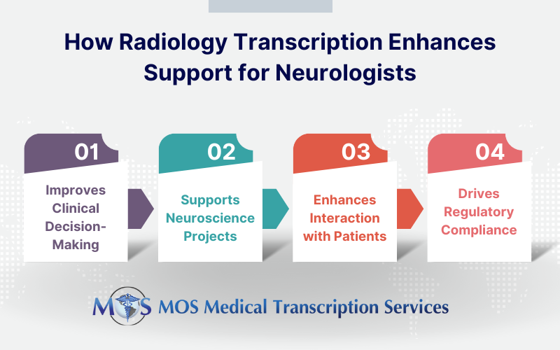
How Radiology Transcription supports Neurologists and Researchers
- Improving Clinical Decision-Making: By providing exact and in-depth radiological reports, transcription plays a critical role in assisting neurologists in correctly diagnosing and treating neurological diseases. By ensuring accurate MRI, CT scan, and other imaging investigation reports, radiology transcription guides neurologists’ treatment decisions and supports proper care delivery.
- Supporting Neuroscience Projects: Radiology transcription is crucial to preserving the precision and dependability of imaging data in neurological research. Well-documented radiological reports are essential for the analysis of illness processes, treatment evaluations, and biomarker identification by researchers. High-quality transcripts ensure the consistency and reproducibility of research data, supporting neurological research projects.
- Enhancing Interaction and Treatment with Patients: Precise transcriptions of radiology tests facilitate clear documentation of imaging findings, which improves communication between neurologists and patients. Patients gain a better understanding of their diagnosis and available treatments as a result. Furthermore, accurate transcriptions guarantee that all members of the medical team are on the same page, which facilitates coordinated and efficient care-especially when it comes to the management of long-term neurological disorders.
- Encouraging Compliance with Laws and Ethics: Maintaining comprehensive medical records is necessary for both clinical practice and research, as well as for legal and ethical compliance. This is where transcription comes in. Precise documentation upholds the validity of the findings and protects medical professionals and investigators from legal disputes.
Different Sections in a Transcribed Radiology Report
- The particulars of the imaging study: Here the types of images taken, contrast material or medications used, and any relevant conditions related to the surgery (if the imaging is done in an operating room) are mentioned. For e.g.Procedure: MRI of the brain without contrast
- In case the results need to be compared with those of an earlier study, the date and name of the comparison study will also be provided in this section or beneath a Comparison Heading.
- Why the study was performed: This section could be titled History, Indication, or Clinical History. The information given here would include the reason for the study (for e.g. chronic headache), and clinical history (for instance, “the patient is a 50-year-old woman with chronic headaches”).
- The technique employed: Here you will have a description of how the procedure was performed, and the kind of images obtained. For simple procedures, the description would be simple and for complex procedures a more detailed step-by-step description would be provided.
- Results: This section would include all values, measurements, and observations recorded during the study. This section may also be titled as Findings or Interpretation. Here, any incidental findings that may be unrelated to the condition for which the study was performed, will also be recorded.
- Impression: In this part of the report, the radiologist would provide his/her assessment of the significance of the findings. Specific diagnoses or suggestions of a probable diagnosis may be given.
Neurologists and other healthcare professionals can benefit by partnering with a HIPAA-compliant medical transcription company specialized in radiology transcription. Transcriptionists in such companies understand the specific terminology related to neurological diagnoses and procedures, ensuring the accuracy of transcripts for neurology and any related radiology reports.